An Overview of the Human Nervous System
A complete guide to the human nervous system — neurons, CNS, PNS, how nerve signals travel, the brain's key regions, and the autonomic nervous system.
A complete guide to the human nervous system — neurons, CNS, PNS, how nerve signals travel, the brain's key regions, and the autonomic nervous system.
You are reading these words right now because of an almost incomprehensibly complex network of communication. This network processes light from this screen, decodes the shapes of letters into language, and retrieves memories of what those words mean.
Simultaneously, it manages your heartbeat, your breathing, your posture, and probably a background hum of thoughts that have nothing to do with biology. All of this, every moment of every day, is the work of your nervous system.
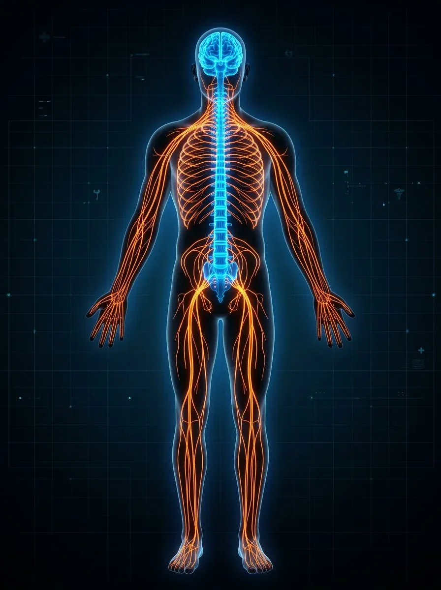
The human nervous system is the body’s command and control network. It collects information from the environment and from the body itself, processes that information at extraordinary speed, and generates responses — from the deliberate (deciding to pick up a cup) to the reflexive (pulling your hand away from a hot surface before you’ve consciously registered the pain). It gives rise to perception, emotion, memory, language, and consciousness itself.
Exploring the architecture of the human nervous system from the top down reveals its two main divisions, the remarkable cells that make it work, how electrical and chemical signals travel between those cells, the organization of the brain, the spinal cord, and the body-wide network of peripheral nerves that carries information to and from every organ and tissue.
The human nervous system is conventionally divided into two main parts:
These divisions are anatomically distinct but functionally inseparable. The CNS processes information and generates commands; the PNS carries sensory information to the CNS and motor commands from the CNS to the muscles and organs.
The brain and spinal cord are the most protected organs in the body. The skull and vertebral column encase them in bone, and a set of three membranes — the meninges — wrap around them, with a layer of cerebrospinal fluid (CSF) between the inner two membranes providing additional cushioning and removing metabolic waste. The blood-brain barrier, a specialised lining of the brain’s blood vessels, restricts which molecules can enter the neural tissue, protecting it from many pathogens and toxins.
The PNS is itself divided into two components:
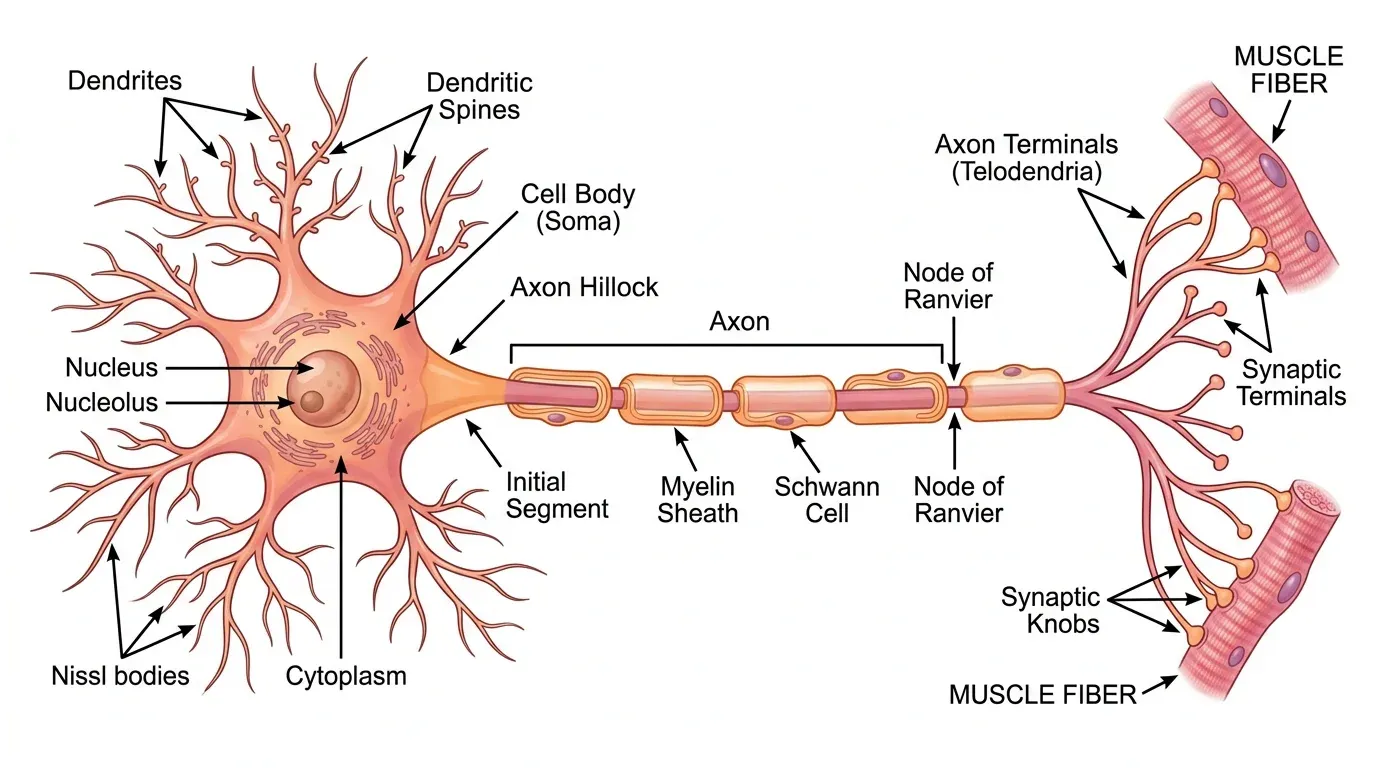
The basic cellular unit of the nervous system is the neuron, or nerve cell. The human brain alone contains an estimated 86 billion neurons. Each neuron is a marvel of biological engineering — a cell specialised to receive, process, and transmit electrical signals. These biological neurons has inspired the artificial neural network that we all are aware of in this age of AI.
A typical neuron has three main parts:
Many axons are wrapped in a fatty insulating layer called myelin, produced by specialised support cells called Schwann cells (in the PNS) or oligodendrocytes (in the CNS). Myelin dramatically speeds up signal transmission and gives the “white matter” of the brain its characteristic colour.
Beyond neurons, the nervous system contains roughly equal numbers of glial cells (sometimes called neuroglia), which provide structural support, produce myelin, regulate the chemical environment around neurons, and play active roles in information processing. Far from being passive scaffolding, glial cells — particularly astrocytes and microglia — are increasingly understood as active participants in brain function and disease.
The currency of communication in the nervous system is the action potential — a rapid, self-propagating electrical impulse that travels along the axon. To understand it, we first need to understand the resting state of a neuron.
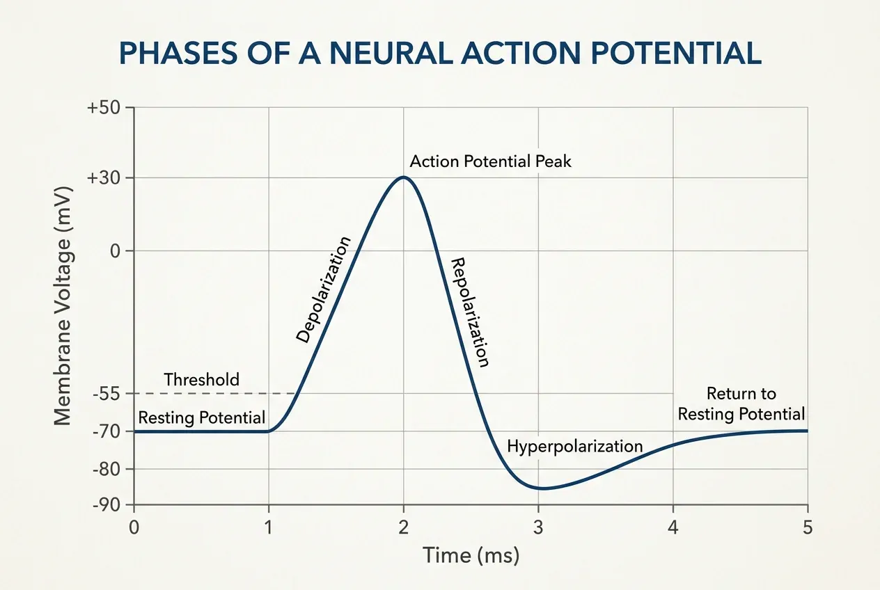
A resting neuron maintains an electrical imbalance across its cell membrane. The inside of the cell is negatively charged relative to the outside, typically around –70 millivolts (mV). This resting membrane potential is maintained by ion pumps and channels — particularly the sodium-potassium pump, which continuously moves sodium ions (Na⁺) out of the cell and potassium ions (K⁺) in, using energy from ATP.
When a neuron receives enough stimulation from its dendrites, the membrane potential at the axon hillock — the junction between cell body and axon — depolarizes toward a threshold of roughly –55 mV. If it reaches this threshold, voltage-gated sodium channels in the membrane snap open. This allows a flood of sodium ions (Na⁺) to rush into the cell. As a result, the membrane potential shoots up to about +40 mV in less than a millisecond.
This rapid depolarization triggers the same process in the adjacent segment of the axon membrane, and so the action potential propagates. It does not travel like electricity through a wire, but rather like a wave of opening and closing ion channels traveling along the axon. The action potential is all-or-nothing: either the threshold is reached and a full-sized impulse fires, or it doesn’t fire at all.
In myelinated axons, the action potential jumps between the gaps in the myelin sheath (nodes of Ranvier) in a process called saltatory conduction, which can achieve speeds of up to 120 metres per second.
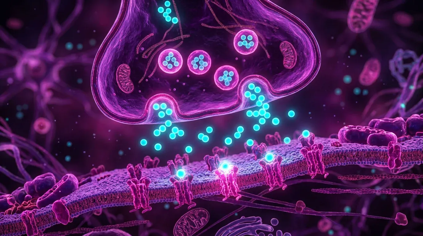
When an action potential reaches the end of an axon — the axon terminal — it triggers the release of chemical messengers called neurotransmitters. These are stored in tiny membrane-bound sacs called synaptic vesicles and are released into the narrow gap between neurons called the synapse (or synaptic cleft).
Neurotransmitters diffuse across the cleft and bind to receptor proteins on the next neuron’s dendrites or cell body. Depending on the neurotransmitter and receptor involved, this can either excite the next neuron (pushing it toward threshold) or inhibit it (pushing it away from threshold). Major neurotransmitters include glutamate (the most common excitatory neurotransmitter in the brain), GABA (the main inhibitory one), dopamine, serotonin, and acetylcholine.
The human brain has an estimated 100–500 trillion synapses. The pattern of their connections — and their strengthening or weakening over time in response to experience — is believed to be the physical basis of memory and learning.
[!NOTE] Multiple sclerosis (MS) is a disease in which the immune system attacks the myelin sheath around nerve fibres in the CNS, disrupting signal transmission. Depending on which fibres are affected, symptoms can include vision problems, muscle weakness, coordination difficulties, and fatigue — reflecting the disruption of normally rapid, precise signal propagation.
The human brain weighs about 1.4 kilograms and contains roughly 86 billion neurons, each potentially connected to thousands of others. Understanding exactly how the brain works begins with its anatomy: its major regions each have distinct, though overlapping, functions.
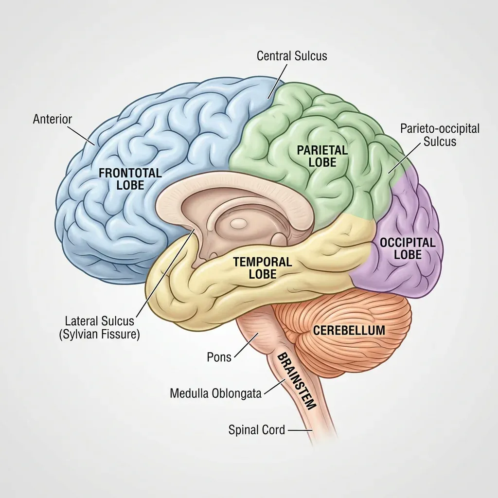
The cerebrum is the largest part of the brain, divided into two hemispheres (left and right) connected by a thick bundle of nerve fibres called the corpus callosum. The outer layer of the cerebrum — the cerebral cortex — is a 2–4 mm layer of grey matter that is heavily folded (the folds are called gyri; the grooves between them are called sulci), greatly increasing its surface area. The cortex is responsible for higher cognitive functions: perception, voluntary movement, language, reasoning, and consciousness.
The cerebrum is divided into four lobes:
Tucked beneath the back of the cerebrum, the cerebellum (Latin for “little brain”) coordinates movement, balance, and fine motor control. It receives signals from the motor cortex (what the brain intended to do) and from proprioceptors in the muscles and joints (what the body is actually doing), and continuously adjusts motor commands to keep movement smooth and coordinated. Damage to the cerebellum produces characteristic movement disorders — stumbling gait, tremor, difficulty performing precise movements — without causing paralysis.
The brainstem connects the cerebrum and cerebellum to the spinal cord and controls essential life functions. It comprises three regions:
The limbic system is a set of structures at the inner edge of the cortex involved in emotion, motivation, and memory. Key components include the hippocampus (essential for forming new long-term memories), the amygdala (central to processing fear, aggression, and other strong emotions), and the hypothalamus (which regulates hunger, thirst, body temperature, and hormonal output from the pituitary gland).
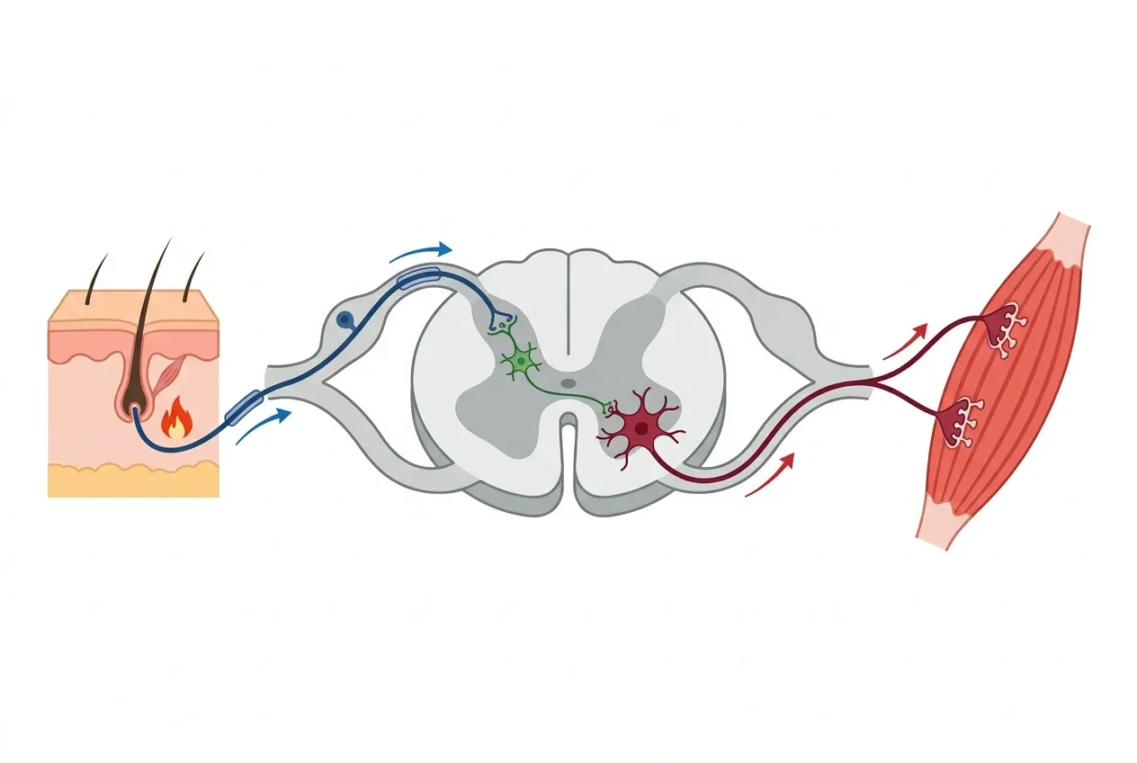
The spinal cord is a column of nervous tissue running from the base of the brain down through the vertebral column, ending around the first or second lumbar vertebra. It performs two main functions: relaying signals between the brain and the body, and integrating certain reflexes without involving the brain at all.
Spinal reflexes — like the knee-jerk reflex, or snatching your hand away from a hot object — involve a sensory signal travelling into the spinal cord, a direct synaptic connection to a motor neuron, and a motor command going out to the muscle, all without the signal having to travel up to the brain and back. This saves critical milliseconds when fast protective responses are needed.
The autonomic nervous system (ANS) regulates the body’s internal environment — the so-called visceral functions — largely outside conscious awareness. It consists of two divisions with complementary effects:
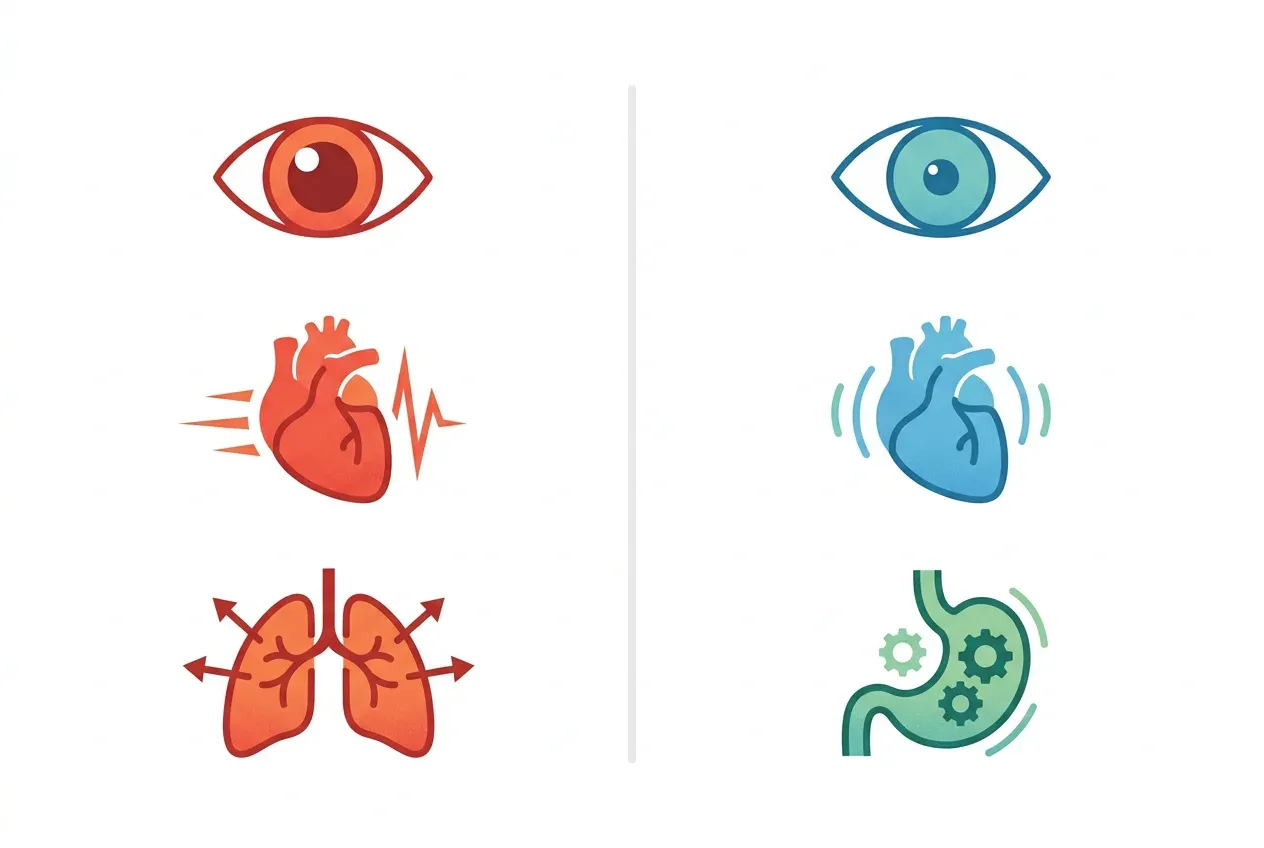
The sympathetic division prepares the body for vigorous physical activity — the classic “fight or flight” response. When activated, it increases heart rate and blood pressure, dilates airways to allow more oxygen uptake, redirects blood from the gut to the muscles, dilates the pupils, and triggers the release of adrenaline (epinephrine) from the adrenal glands. Digestion slows; energy reserves are mobilised. In ancestral environments, this response was triggered by predators; today, it is also triggered by stress, anxiety, and perceived social threats.
The parasympathetic division promotes restoration and digestion — the “rest and digest” state. It slows the heart rate, stimulates digestive activity, promotes gland secretion, and constricts the pupils. The vagus nerve, the longest parasympathetic nerve, connects the brainstem to the heart, lungs, and most abdominal organs, influencing them all.
The two divisions are generally (though not always) antagonistic, and their balance is continuously adjusted by the hypothalamus in response to internal and external conditions.
The central nervous system (CNS) consists of the brain and spinal cord — the processing and command centres. The peripheral nervous system (PNS) includes all the nerves and ganglia outside the CNS, carrying sensory information to the brain and motor commands from the brain to muscles and organs. The somatic PNS handles voluntary movement and sensation; the autonomic PNS handles involuntary functions like heart rate and digestion.
A neuron is a specialised cell that transmits electrical and chemical signals. It has three main parts: dendrites that receive signals from other neurons or sensory receptors, a cell body (soma) that processes those signals, and an axon that carries the output signal to the next cell. Neurons communicate at junctions called synapses by releasing chemical messengers called neurotransmitters.
An action potential is a rapid, self-propagating electrical impulse that travels along a neuron's axon. It is triggered when incoming signals depolarise the membrane to a threshold (around –55 mV), causing voltage-gated sodium channels to open and sodium ions to rush into the cell, briefly reversing the charge across the membrane. This process sweeps along the axon to the next synapse, where it triggers the release of neurotransmitters.
The cerebrum (divided into frontal, parietal, temporal, and occipital lobes) handles higher functions: thought, voluntary movement, language, perception, and memory. The cerebellum coordinates movement and balance. The brainstem (midbrain, pons, and medulla oblongata) controls vital autonomic functions — breathing, heart rate — and relays information between the brain and spinal cord. The limbic system (including the hippocampus and amygdala) is involved in emotion, memory, and motivation.
The fight-or-flight response is the activation of the sympathetic division of the autonomic nervous system in response to perceived threat or stress. It raises heart rate and blood pressure, dilates airways and pupils, redirects blood to skeletal muscles, and stimulates adrenaline release — all preparing the body for rapid physical action. The opposite parasympathetic state — rest and digest — promotes recovery, digestion, and restoration when the threat has passed.
Applaud the article to let us know. You can clap multiple times!